Carotid and Vertebral Duplex Ultrasound
 Unfortunately, the great majority of individuals that experience a stroke, one of the most feared cardiovascular events, have no warning symptoms prior to their event. The Carotid and Vertebral Duplex Ultrasound evaluates the overall structure and health of the carotid and vertebral arteries. These arteries are located on either side of the neck, and are tasked with the responsibility of carrying blood to the brain. These arteries may accumulate of plaque, or “atherosclerosis”, which is an indicator of the potential for stroke, as well as a marker for overall cardiovascular health.
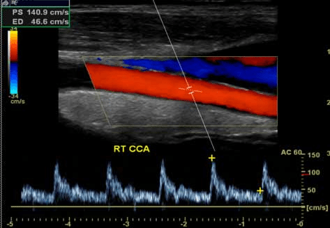
The Carotid and Vertebral Duplex Ultrasound test utilizes high frequency ultrasound to create a visualization (like the one pictured below) of the blood vessel walls and blood flow within the carotid and vertebral arteries. Ultrasound waves are sent through a device called a transducer to evaluate the carotid and vertebral arteries, looking at the structure and function of the vessels to detect plaque build-up and blood flow disturbances. This allows Dr. Marcus to determine the health of these vital arteries.
The results of a Carotid and Vertebral Duplex Ultrasound will immediately provide valuable information on the status of these critical vessels, their level of plaque build up, and thickness of their lining.
A thorough evaluation of the carotid and vertebral arteries allows Dr. Marcus and his patients to implement a plan designed specifically for each patient’s individual risk. This can help ensure that the proper measures are taken to treat any abnormalities that are discovered, and to prevent the development and progression of disease.